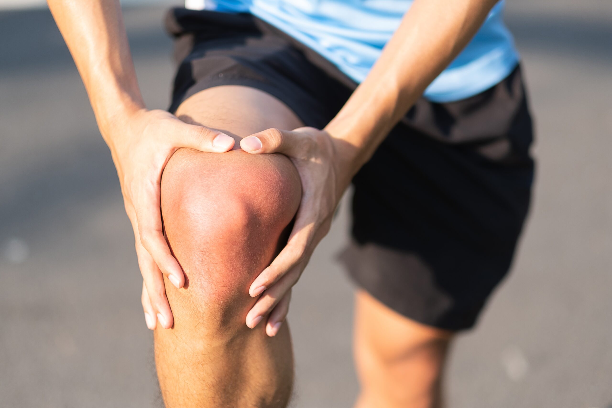
Patellar instability means that the kneecap (patella) does not glide stably in its groove and partially or completely jumps out of the joint towards the outside (patellar dislocation). This problem frequently affects young, active people and athletes, but can in principle occur at any age. Thanks to new scientific findings and modern surgical techniques, very good long-term stability of the knee can now be achieved for most patients.
How does a patellar dislocation occur?
The first dislocation of the kneecap often results from a sports injury, for example while playing football, skiing, playing handball or during rapid changes of direction in sport. The dislocation almost always involves an injury to an important ligament: the medial patellofemoral ligament (MPFL). This ligament is the most important stabiliser of the kneecap towards the inside. If it tears or is overstretched, the patella loses its guidance and the risk of further dislocations rises considerably. Early and correct initial treatment is therefore crucial, as repeated dislocations can lead to cartilage damage and osteoarthritis in the long term.
Typical symptoms
Typical symptoms of patellar instability are the sudden popping out of the kneecap, severe knee pain and swelling of the knee joint. Many patients also report that the kneecap “shifts” or “feels insecure”. This often leads to a constant feeling of instability, a great fear of another dislocation and reduced resilience during sport.
Anatomical risk factors
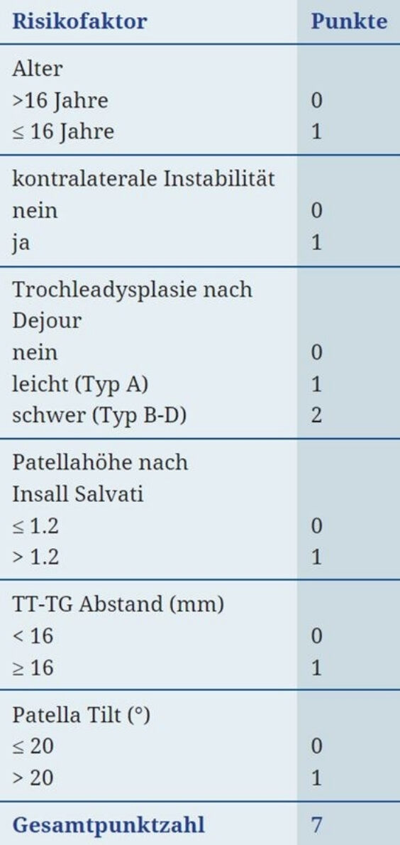
In addition to the accident mechanism, there is often an anatomical predisposition that reduces the stability of the kneecap. These factors, which are examined closely during the assessment, include rotational malalignments of the thigh bone (femur) or shin bone (tibia). Such malalignments can cause the patella to be pulled more strongly towards the outside. This is assessed with special CT or MRI measurements such as the TT-TG distance, which should ideally be below 16 millimetres.
A malalignment of the leg axis, such as knock knees, or a flat/pronated foot also increases the outward pull. The shape of the bony groove (trochlea) also plays an important role. If this groove is flat or convex instead of a stabilising concave shape, this is known as trochlear dysplasia. Finally, a kneecap that sits too high (patella alta) also increases the risk, as it has less bony guidance.
Risk analysis after dislocation
After every patellar dislocation, a precise risk analysis should be carried out. The Patellar Instability Severity Score (PIS score) is frequently used for this purpose. It evaluates various factors such as age, anatomical risk factors, the height of the patella, possible trochlear dysplasia and the TT-TG distance. The score helps the doctor decide whether conservative treatment is sufficient or whether surgery is advisable.
Conservative treatment and physiotherapy
After a first dislocation, conservative treatment without surgery can often be carried out initially. The goals are healing of the MPFL ligament, stabilisation through muscle training and restoration of leg axis control. At the beginning, a patella-centring knee brace is usually worn to stabilise the kneecap during the healing phase.
A central component of the therapy is physiotherapy with targeted muscle training for the inner part of the quadriceps (M. vastus medialis), the core muscles, the hip abductors and the foot muscles. The range of motion (flexion) is increased step by step according to the following typical scheme:
| Period | Range of motion (flexion) |
|---|---|
| Week 1–2 | 0–20–40° |
| Week 3–4 | 0–10–60° |
| Week 5–6 | 0–0–90° |
In addition, partial weight-bearing is usually required for about two weeks. In the case of leg axis malalignment, stabilising insoles can also help.
When is surgery advisable?
Surgery may be recommended in the case of repeated dislocations, a high risk of re-dislocation, significant anatomical malalignments or if cartilage and bone injuries are already present. Depending on the cause and the individual anatomy of the patient, various modern procedures are available. These include MPFL reconstruction, tibial tubercle transfer, trochleoplasty or, in rare cases, an axis-correcting osteotomy.
Key points to remember
The stability of the kneecap depends largely on the bone shape, the ligament structures and the leg axis. Every patellar dislocation should be assessed with X-ray and MRI in order to estimate the risk of further dislocations using the PIS score. Correctly performed follow-up treatment is in any case crucial for long-term success.
Frequently asked questions about patellar instability
No. After a first dislocation, conservative treatment is often possible initially – with a patella-centring knee brace, physiotherapy and targeted muscle training. The PIS score helps to assess whether the risk of re-dislocation makes surgery advisable.
The medial patellofemoral ligament (MPFL) is the most important stabiliser of the kneecap towards the inside. It is almost always injured during a dislocation. If it tears or is overstretched, the patella loses its guidance and the risk of further dislocations rises considerably.
Rotational malalignments of the thigh or shin bone, an increased TT-TG distance, knock knees or a flat/pronated foot, trochlear dysplasia and a kneecap that sits too high (patella alta).
In the case of repeated dislocations, a high risk of re-dislocation, significant anatomical malalignments or existing cartilage and bone injuries. Depending on the anatomy, MPFL reconstruction, tibial tubercle transfer, trochleoplasty or, rarely, an axis-correcting osteotomy may be considered.
Repeated dislocations can lead to cartilage damage and osteoarthritis in the long term. Every patellar dislocation should therefore be assessed with X-ray and MRI and the risk of further dislocations estimated.