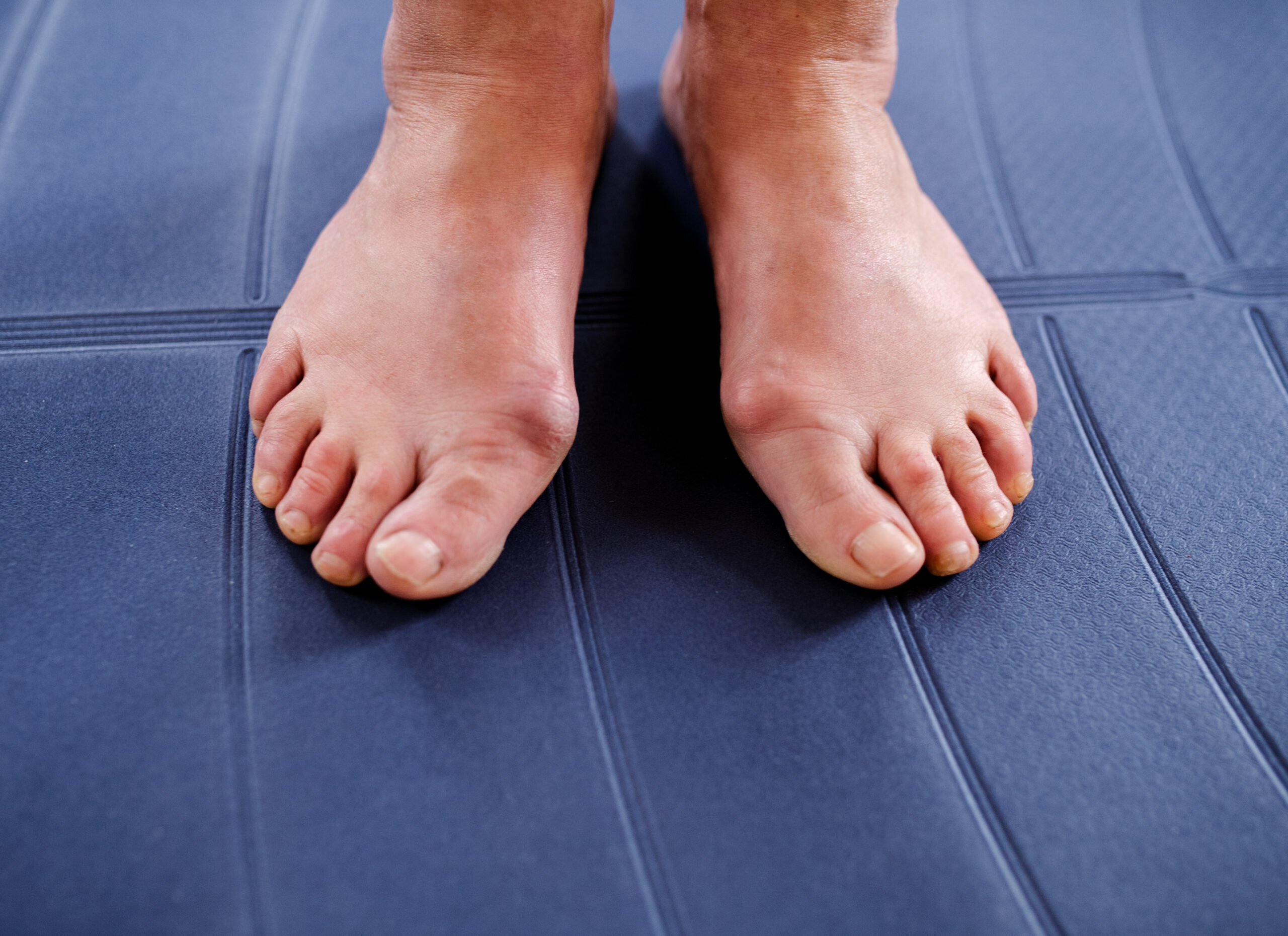
Splayfoot is one of the most common acquired forefoot deformities in adulthood and, in orthopaedic surgical practice, a central starting point for numerous further deformities. Contrary to common belief, it is not merely a functional or cosmetic problem but a biomechanically relevant instability of the forefoot which, if left untreated, can lead to chronic pain, gait disorders and complex secondary deformities.
1. What is a splayfoot?
In a healthy foot, the five metatarsal bones form a stable transverse arch. The main load is distributed across three defined points: the heel, the first and the fifth ray. In a splayfoot, the metatarsal bones drift apart – particularly the central rays (II–IV) – and the transverse arch collapses.
The result is a pathological shift of pressure onto structures that are not designed for sustained loading. This altered statics forms the basis for pain (metatarsalgia) and progressive forefoot deformities.
2. Causes and pathomechanics
In most cases, splayfoot is acquired. Common causes include:
- Ligament and connective tissue insufficiency (age-related or genetic)
- Muscular imbalance of the intrinsic foot muscles
- Chronic overload (prolonged standing, excess weight)
- Unphysiological footwear (high heels, narrow toe box)
With the loss of transverse arch stability, a biomechanical chain reaction begins: the first ray loses its load-bearing function, the second and third rays take on a disproportionate share of the load, and the forefoot as a whole becomes wider.
3. Splayfoot as a cause of hallux valgus and tailor's bunion
Clinical practice shows that splayfoot is frequently the mechanical basis for the development of further forefoot deformities:
- Hallux valgus: due to the instability of the transverse arch, the first metatarsal drifts medially while the big toe deviates laterally.
- Digitus quintus varus (tailor's bunion): the widening of the forefoot pushes the fifth ray outwards, creating a painful prominence.
4. Symptoms and clinical warning signs
- Load-dependent pain beneath the metatarsal heads (metatarsalgia)
- Calluses, corns, a painful “worn-through” fat pad
- An increasingly wide forefoot with growing shoe conflicts
- Altered roll-over pattern and gait
- Combination with hammer or claw toes
Late sequelae of untreated splayfeet can include Morton's neuromas, transfer metatarsalgia after previous surgery and degenerative changes in the Lisfranc joint.
5. Diagnostics from a surgical perspective
Precise diagnostics are decisive for surgical planning:
- Clinical examination while standing and walking
- Weight-bearing X-rays of the forefoot to assess ray lengths and axes
- Pedobarography for objective pressure analysis
- Assessment of accompanying deformities (hallux valgus, digitus quintus varus, toe deformities)
6. Conservative treatment – recognising its limits
Insoles with metatarsal pads, shoe modifications and targeted foot exercises can relieve symptoms and slow progression. An anatomical correction of the splayfoot, however, cannot be achieved conservatively.
If pain persists or deformities progress, surgical evaluation should take place early in order to avoid complex secondary damage.
7. Surgical treatment of splayfoot
When is surgery advisable?
- Persistent metatarsalgia despite adequate conservative treatment
- Progressive forefoot widening with shoe conflict
- Combined deformities (hallux valgus, digitus quintus varus)
- Recurrent symptoms after isolated toe surgery
The basic surgical principle
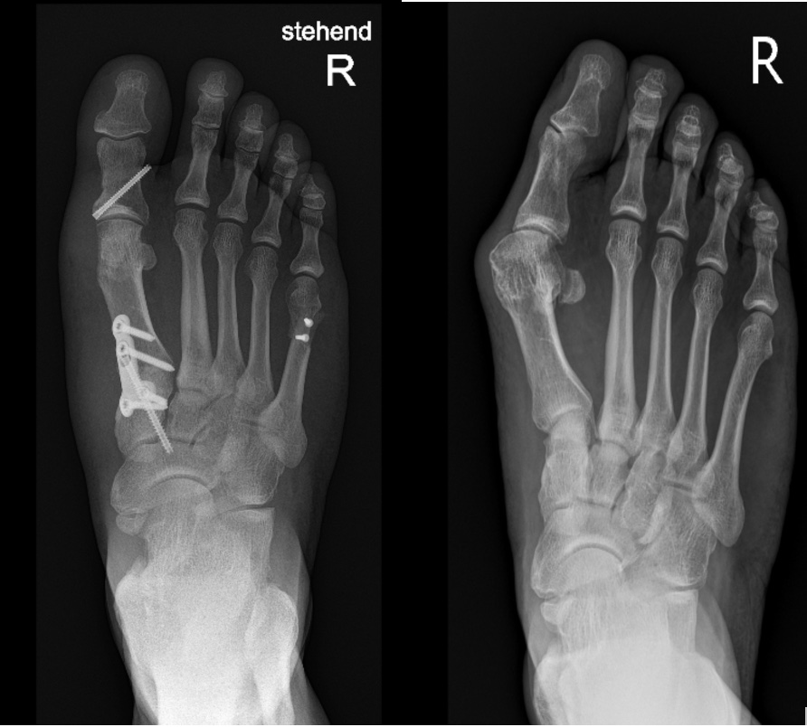
The aim of surgery is to restore functional forefoot statics. As a rule, this can only be achieved through combined procedures.
The Weil osteotomy
The Weil osteotomy is an established standard procedure for treating splayfoot-related metatarsalgia:
- Targeted division and proximal shift of the overloaded metatarsal bone
- Pressure relief of the painful metatarsal heads
- Fixation with small screws
Combined procedures
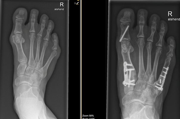
In many cases, the Weil osteotomy is combined with further corrections:
- Surgical hallux valgus correction
- Correction of digitus quintus varus
- Tendon-balancing procedures on the toes
Only this holistic forefoot correction achieves a permanently stable and pain-free result.
8. Aftercare and prognosis
Postoperatively, mobilisation takes place in a forefoot off-loading shoe for about 4–6 weeks. Full weight-bearing is built up step by step. As a rule, a return to sporting activities is possible after around three months.
With the correct indication, the results of modern surgical techniques are very good — particularly when the splayfoot, as the cause of the overall problem, is consistently treated as part of the procedure.
Frequently asked questions about splayfoot
No. An acquired splayfoot does not regress on its own. Insoles, shoe modifications and foot exercises can relieve symptoms and slow progression, but the deformity itself cannot be corrected conservatively.
When load-dependent pain beneath the metatarsal heads persists despite consistent conservative treatment, the forefoot keeps widening, or secondary deformities such as hallux valgus and tailor's bunion develop. Early surgical evaluation helps to avoid complex secondary damage.
An established standard procedure for splayfoot-related metatarsalgia: the overloaded metatarsal bone is divided in a targeted manner, shifted back and fixed with small screws, relieving pressure on the painful metatarsal heads.
It is possible, but isolated toe corrections that ignore the underlying splayfoot frequently lead to recurrence. Sustainable results require a holistic concept that also corrects the disturbed forefoot statics.
Mobilisation takes place in a forefoot off-loading shoe for about 4–6 weeks, after which weight-bearing is increased step by step. A return to sport is usually possible after around three months.
Where there is a medical indication, surgical correction of splayfoot and its secondary deformities is covered by Swiss basic health insurance. We clarify coverage with you before the procedure.